Anatomy Of Chest - Chest Wall Anatomy Images Stock Photos Vectors Shutterstock. Angina is the term for chest pain caused by poor blood flow to the heart. Get the full built by science program: The circulatory system does most of its work. Related posts of anatomy of the chest and stomach abdominal muscles picture anatomy. Related posts of anatomy of the chest area anatomy of penis.
It provides protection to vital organs (eg, heart and major vessels, lungs, liver) and provides stability for movement. The muscles of the chest develop from the somites found in the mesoderm. Pacemaker diagram cross section of a human heart with pacemaker fitted, showing the major arteries and veins. Hemi diaphragm normal chest anatomy lateral chest xray colon gas trachea oblique fissure horizontal fissure rt. Angina is the term for chest pain caused by poor blood flow to the heart.
Lateral Anatomy Of The Chest Bones And Abdomen Medical Art Works from cdn.shopify.com Anatomy of the chest, abdomen, and pelvis was produced in part due to the generous funding of the david f. The epidermis is the outermost layer that provides a protective, waterproof seal over the body. It's also sometimes referred to as the breastbone. The pig has a large horn plate, perforated by the two nares. It provides protection to vital organs (eg, heart and major vessels, lungs, liver) and provides stability for movement. (1) the pectoralis major, and (2) the pectoralis minor. Diseases of the chest and chest abnormalities make up a significant portion of a physician's daily practice. The circulatory system does most of its work.
Chest a man's chest — like the rest of his body — is covered with skin that has two layers.
In insects, crustaceans, and the extinct trilobites, the thorax is one of the three main divisions of the creature's body, each of which is in turn composed of multiple segments. Swensen fund for innovation in teaching. The muscles of the chest develop from the somites found in the mesoderm. The pectoralis major, pectoralis minor, serratus anterior and subclavius. It contains four muscles that exert a force on the upper limb: Computed tomography (ct) of the chest can detect pathology that may not show up on a conventional chest radiograph(1). Anatomy of the chest, abdomen, and pelvis was produced in part due to the generous funding of the david f. It is not unusual to have areas of bruising or erosion to the dorsal tip of the rostal plane. The chest anatomy includes the pectoralis major, pectoralis minor and the serratus anterior. A heart attack results from blocked blood flow, often from a blood clot, to your heart muscle. The chest or thorax is the region between the neck and diaphragm that encloses organs, such as the heart, lungs, esophagus, trachea, and thoracic diaphragm. This is an eps 10 vector illustration and includes a high resolution jpeg. It is important to remember the position and orientation of the heart when placing a stethoscope on the chest of a patient and listening for heart sounds, and also when looking at images taken from a midsagittal perspective.
Browse 6,406 chest anatomy stock photos and images available, or search for human anatomy to find more great stock photos and pictures. This tutorial is designed to help you understand the normal anatomy of the chest as seen on ct images in three planes: Anatomy of the thorax, heart, abdomen and pelvis recommended text gray's anatomy for students. In insects, crustaceans, and the extinct trilobites, the thorax is one of the three main divisions of the creature's body, each of which is in turn composed of multiple segments. The chest anatomy includes the pectoralis major, pectoralis minor and the serratus anterior.
Figure 2 From Introduction To Chest Wall Reconstruction Anatomy And Physiology Of The Chest And Indications For Chest Wall Reconstruction Semantic Scholar from d3i71xaburhd42.cloudfront.net Here, we break down the anatomy of your chest muscles. The chest wall is comprised of skin, fat, muscles, and the thoracic skeleton. The circulatory system does most of its work. Chest a man's chest — like the rest of his body — is covered with skin that has two layers. Chest pain has many possible causes, all of which need medical attention. Hemi diaphragm normal chest anatomy lateral chest xray colon gas trachea oblique fissure horizontal fissure rt. It is not unusual to have areas of bruising or erosion to the dorsal tip of the rostal plane. It's also sometimes referred to as the breastbone.
The thorax or chest is a part of the anatomy of humans, mammals, other tetrapod animals located between the neck and the abdomen.
The chest is made up primarily of two muscles: Angina is the term for chest pain caused by poor blood flow to the heart. The clinical anatomy of the respiratory tract starts at the external nares or nose. Your sternum protects the organs of your torso from injury and also serves as a. It spreads out like a fan and covers the rib cage like an armor plate. Get the full built by science program: Chest muscles anatomy (1) pectoralis major muscle. Radiology basics of chest ct anatomy with annotated coronal images and scrollable axial images to help medical students and junior doctors learning anatomy. Computed tomography (ct) of the chest can detect pathology that may not show up on a conventional chest radiograph(1). Related posts of anatomy of the chest area anatomy of penis. This page provides an overview of the chest muscle group. Browse 2,553 female chest anatomy stock photos and images available, or start a new search to explore more stock photos and images. A heart attack results from blocked blood flow, often from a blood clot, to your heart muscle.
Related posts of anatomy of the chest area anatomy of penis. The chest or thorax is the region between the neck and diaphragm that encloses organs, such as the heart, lungs, esophagus, trachea, and thoracic diaphragm. It is enclosed by the ribs, the vertebral column, and the sternum, or breastbone, and is separated from the abdominal cavity (the body's largest hollow space) by a muscular and membranous partition, the diaphragm. Diseases of the chest and chest abnormalities make up a significant portion of a physician's daily practice. Learn about each of these muscles, their locations, functional anatomy and exercises for them.
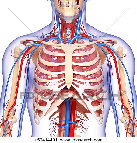
Chest Anatomy Artwork Clip Art U59414401 Fotosearch from fscomps.fotosearch.com Chest pain has many possible causes, all of which need medical attention. Here's how science can help you grow! Thoracic cavity, also called chest cavity, the second largest hollow space of the body. The right side of the heart is deflected anteriorly, and the left side is deflected posteriorly. Radiology basics of chest ct anatomy with annotated coronal images and scrollable axial images to help medical students and junior doctors learning anatomy. It provides protection to vital organs (eg, heart and major vessels, lungs, liver) and provides stability for movement. The chest is made up primarily of two muscles: The pectoralis major and the pectoralis minor, known collectively as your pecs.
Pacemaker diagram cross section of a human heart with pacemaker fitted, showing the major arteries and veins.
Radiology basics of chest ct anatomy with annotated coronal images and scrollable axial images to help medical students and junior doctors learning anatomy. A heart attack results from blocked blood flow, often from a blood clot, to your heart muscle. Muscles of the chest and their functions you have two mighty muscles on both sides of your chest: Get the full built by science program: The first step in understanding thorax anatomy is to find out its boundaries. Chest muscles anatomy (1) pectoralis major muscle. It provides protection to vital organs (eg, heart and major vessels, lungs, liver) and provides stability for movement. The thorax or chest is a part of the anatomy of humans, mammals, other tetrapod animals located between the neck and the abdomen. It's also sometimes referred to as the breastbone. Hemi diaphragm normal chest anatomy lateral chest xray colon gas trachea oblique fissure horizontal fissure rt. Chest pain has many possible causes, all of which need medical attention. The myotomes elongate and invade the mesoderm of the wall of the embryonic thoracic and abdominal cavities. It spreads out like a fan and covers the rib cage like an armor plate.
Share :
Post a Comment
for "Anatomy Of Chest - Chest Wall Anatomy Images Stock Photos Vectors Shutterstock"


Post a Comment for "Anatomy Of Chest - Chest Wall Anatomy Images Stock Photos Vectors Shutterstock"